Shop Amazon - Up to 25% off pre-owned iPhones
If you really want some breakthrough in Artificial intelligence you require to look at Bio chemistry in human and the coordination in the human body like ATP Hydrolysis and how it helps in contraction of Muscles Fibers in human,its basic building block called monomers which we can define programmatically as class and its repetition and coordination with external and internal environment to develop some effective system which we call Programs.One of the area from where Artificial Intelligence in Machine Learning has been reprieved is a Neural Network which Exist in the human neural system.
NEURAL SYSTEM:
The neural system of all animals is composed of highly specialised cells called neurons which can detect, receive and transmit different kinds of stimuli.The neural organisation is very simple in lower invertebrates. For example, in Hydra it is composed of a network of neurons. The neural system is better organised in insects, where a brain is present along with a number of ganglia and neural tissues. The vertebrates have a more developed neural system.In terms of Artificial Intellegence.A neuron is a cell in the brain whose principal function is to collect is the collection,processing,dissemination of electrical signal just like a small class in a Data structure.The brain's information processing capacity is thought to emerge mainly from networks of such class called Neuron.So we can think of Neurons as micro chip which is a class in programming language having Data Structures and ability to work upon information stored in these Data structure thus I called it a class in terms of programming language.These class are Well connected by very complex communication system that we call as Neural Networks.
HUMAN NEURAL SYSTEM
The human neural system is divided into two parts :
(i) the central neural system (CNS)
(ii) the peripheral neural system (PNS)
The CNS includes the brain and the spinal cord and is the site of information processing and control. The PNS comprises of all the nerves of the body associated with the CNS (brain and spinal cord). The nerve fibres of the PNS are of two types :
(a) afferent fibres
(b) efferent fibres
The afferent nerve fibres transmit impulses from tissues/organs to the CNS and the efferent fibres transmit regulatory impulses from the CNS to the concerned peripheral tissues/organs. The PNS is divided into two divisions called somatic neural system and autonomic neural system. The somatic neural system relays impulses from the CNS to skeletal muscles while the autonomic neural system transmits impulses from the CNS to the involuntary organs and smooth muscles of the body. The autonomic neural system is further classified into sympathetic neural system and parasympathetic neural system.
NEURON AS STRUCTURAL AND FUNCTIONAL UNIT OF NEURAL SYSTEM
A neuron is a microscopic structure composed of three major parts,
namely, cell body, dendrites and axon The cell body contains cytoplasm with typical cell organelles and certain granular bodies called Nissl’s granules. Short fibres which branch repeatedly and project out of the cell body also contain Nissl’s granules and are called dendrites.These fibres transmit impulses towards the cell body. The axon is a long fibre, the distal end of which is branched. Each branch terminates as a bulb-like structure called synaptic knob which possess synaptic vesicles containing chemicals called neurotransmitters. The axons transmit nerve impulses away from the cell body to a synapse or to a neuro-muscular junction. Based on the number of axon and dendrites, the neurons are divided into three types, i.e., multipolar (with one axon and two or more dendrites; found in the cerebral cortex), bipolar (with one axon and one dendrite, found in the retina of eye) and unipolar (cell body with one axon only; found usually in the embryonic stage). There are two types of axons, namely, myelinated and nonmyelinated.The myelinated nerve fibres are enveloped with Schwann cells, which form a myelin sheath around the axon. The gaps between two adjacent myelin sheaths are called nodes of Ranvier. Myelinated nerve fibres are found in spinal and cranial nerves. Unmyelinated nerve fibre is enclosed by a Schwann cell that does not form a myelin sheath around the axon, and is commonly found in autonomous and the somatic neural systems.
 Structure of a neuron that can well represented by some Elegant Data structure which i will define in my next page and its functionality receiving,processing , transmitting information and networking will be defined by Methods in a class.The nervous system consists of neurons and supporting cells. Neuron Organization. Neurons and neuroglia are organized into the central nervous system (the brain and spinal cord) and the peripheral nervous system (sensory and motor neurons).
Nerve impulses are produced on the axon membrane.The Resting Membrane Potential. The inside of the membrane is electrically negative in comparison with the
outside.Action Potentials. In response to a stimulus that
depolarizes the membrane, voltage-gated channels open, producing a nerve impulse. One action potential stimulates
the production of the next along the axon.
Neurons form junctions called synapses with other cells. Structure of Synapses. Neurotransmitters diffuse acrossto the postsynaptic cell and combine with receptor proteins. Neurotransmitters and Their Functions. Some neurotransmitters cause a depolarization in the postsynaptic membrane; others produce inhibition by hyperpolarization.
The central nervous system consists of the brain
and spinal cord. The Evolution of the Vertebrate Brain. Vertebrate brains include a forebrain, midbrain, and hind brain. The Human Forebrain. The cerebral cortex contains areas specialized for different functions. The Spinal Cord. Reflex responses and messages to and from the brain are coordinated by the spinal cord.
The peripheral nervous system consists of sensory
and motor neurons. Components of the Peripheral Nervous System. A spinal nerve contains sensory and motor neurons.The Autonomic Nervous System. Sympathetic motor neurons arouse the body for fight or flight; parasympathetic motor neurons have antagonistic actions.
 Three types of neurons. Sensory neurons carry information about the environment to the brain and spinal cord. Association neurons are found in the brain and spinal cord and often provide links between sensory and motor neurons. Motor neurons carry impulses or “commands” to muscles and glands(effectors).
Structure of a typical neuron. Extending from the cell body are many dendrites, which receive information and carry it to the cell body. A single axon transmits impulses away from the cell body. Many axons are encased by a myelin sheath, whose multiple membrane layers facilitate a more rapid conduction of impulses. The sheath is interrupted at regular intervals by small gaps called nodes of Ranvier. In the peripheral nervous system, myelin sheaths are formed by supporting Schwann cells
Mathematically neuron which can then converted to any programming language can be defined by as shown below:

 How mathematically we can define units in neural network that is Neuron.As we know that neural networks are are composed of nodes or units called neuron connected by direct links .A link from neuron j to neuron i serves to propagate the activation aj from node j to i.Each link has a numeric weight Wj,i associated with it.which determines the strength or sign of the connection.Each unit i first computes the weighted sum of its inputs
 are as shown below in the figure


 The figure


The above was the mathematical part which is the part of machine learning based on neural networks i will come to its practical application in data mining,Artificial learning,mean square Error as one of the artificial learning Algorithm etc before going into biology let's see one of its application in communication theory ad adaptive filtering.
ARMA Processes:
An ARMA process for the time series X (n) is given by
 Or in mathematically compressed form as
 Taking the Z-transform of above and solving for the pulse transfer function we get
 which is a filter with both poles and zeros, as shown in Figure below since it is a combination of AR and MA processes. The order of the ARMA process is (p, q).The autocorrelation function of the ARMA (p, q) process, assuming p > q, is
 ARMA filter of order (p, q) with p > q.
LEAST-SQUARE ESTIMATION is also one of the application of Artificial machine Learning and associated to Adaptive filtering .Here we will see that this algorithm is very useful to eliminate noise from a given signal Intelligently
In the least square estimation, the criterion is only to minimize the squared difference between the given data (signal plus noise) and the assumed signal data. Suppose we want to estimate M parameters, denoting the M-dimensional vector θ, from the K measurements, denoting the K-dimensional vector Y with K ≥ M . The relation between the parameters θ and the observed data Y is given by the linear matrix mode
Y = H θ + N
where H is a known (K ×M) matrix, and N is the unknown (K × 1) error vector that occurs in the measurement of θ. The least-square estimator (LSE) of θ chooses the values that make X = H θ closest to the observed data Y. Hence, we minimize

Note that Y T H θ is a scalar. Taking the first-order partial derivative of the cost function J (θ) with respect to θ (i.e., the gradient) and setting it equal to zero, we obtain the set of linear equations
 And Least Square Estimate (LSE) is found to be
 Note that is the Matrix Equation And the inverse of only Square matrix exist if you require to understand it fully join my linear Algebra course.
Note that the second-order partial derivative is
 This matrix is positive-definite as long as H is assumed to be of full rank to guarantee the inversion of HT H Thus, the solution
 is unique and minimizes J (θ) . The equations
 to be solved for θˆ = θˆ ls are referred to as the normal equations We observe that the error in the estimator ls θˆ is a linear function of the measurement errors N, since
 The minimum least-square J min can be shown, after some matrix operation, to be
 Generalization of the Least-Square Problem which finds huge application in machine learning and where we have to optimize certain function H,at certain optimum domain theta.
The least-square cost function can be generalized by introducing a K × K positive definite weighting matrix W to yield
 The elements of the weighting can be chosen to emphasize specific values of the data that are more reliable for the estimate θˆ The general form of the least-square estimator can be shown to be


 W = R−1
RECURSIVE LEAST-SQUARE ESTIMATOR
In real time estimation problems (filtering), it is necessary to write the estimator θˆ in a recursive form for efficiency. For example, consider a situation where an estimate θˆ is determined based on some data YK . If new data YK +1 is to be processed after having determined an estimate based on the data YK , it is best to use the old solution along with the new data to determine the new least-square estimator. It is clear that discarding the estimate based on the data YK and restarting the computation for a solution is inefficient. This procedure of determining the least-square estimate from an estimate based on YK and the new data YK+1 is referred to as sequential least-square estimation, or more commonly recursive least-square (RLS) estimation. Consider the problem of estimating θ from the data vectors ZM given by the linear model
 where
where
 is an (MK +1) collection of vectors Y1 ,Y2 ,K,YM , since each vector Yk , k = 1, 2,K,M, is a (K +1) vector,
 is an (MK +1) error vector, and
 is an (MK × n) mapping matrix relating ZM to the (n×1) parameter vector θ to be estimated.
It can be shown that the RLS estimator is given by
 where
where
 C is the error covariance matrix given by
 And
And
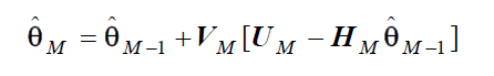
 The covariance matrix of the individual noise vector N is Rii ≜ Ri .The estimator M θˆ based on ZM is formed as a linear combination of θM-1 and a correction term VM [UM − HM θˆ M −1 ] .If θ were a random variable, it can be shown that the generalization of the recursive least-square estimation leads to the Kalman filter i will explain this later. In the next paper on filtering, i will present an introduction to Kalman filtering .
Again come to neural networks


Single Layer Feed Forward Neural network(Perceptrons)
 See above figure.


 This function is linearly seperable.The above figure

 Regression.The Square error for the single training example with input X and true output y mathematically written as
 Where hw(x)=output of the perceptron.
Now the calculus is coming into picture to maximize or minimize certain function at some optimal Domain of the function.


Again coming to biology pare and completing the mechanism of Communication among Neurons and how we can implement that in Program class.
Generation and Conduction of Nerve Impulse:
Neurons are excitable cells because their membranes are in a polarised state. Do you know why the membrane of a neuron is polarised? Different types of ion channels are present on the neural membrane. These ion channels are selectively permeable to different ions. When a neuron is not conducting any impulse, i.e., resting, the axonal membrane is comparatively more permeable to potassium ions (K+) and nearly impermeable to sodium ions (Na+). Similarly, the membrane is impermeable to negatively charged proteins present in the axoplasm. Consequently, the axoplasm inside the axon contains high concentration of K+ and negatively charged proteins and low concentration of Na+. In contrast, the fluid outside the axon contains a low concentration of K+, a high concentration of Na+ and thus form a concentration gradient. These ionic gradients across the resting membrane are maintained by the active transport of ions by the sodium potassium pump which transports 3 Na+ outwards for 2 K+ into the cell. As a result, the outer surface of the axonal membrane possesses a positive charge while its inner surface

Diagrammatic representation of impulse conduction through an axon (at points A and B) becomes negatively charged and therefore is polarised. The electrical potential difference across the resting plasma membrane is called as the resting potential.
You might be curious to know about the mechanisms of generation of nerve impulse and its conduction along an axon. When a stimulus is applied at a site as shoen in the above figure point A on the polarised membrane the membrane at the site A becomes freely permeable to Na+. This leads to a rapid influx of Na+ followed by the reversal of the polarity at that site, i.e., the outer surface of the membrane becomes negatively charged and the inner side becomes positively charged. The polarity of the membrane at the site A is thus reversed and hence depolarized. The electrical potential difference across the plasma membrane at the site A is called the action potential, which is in fact termed as a nerve impulse. At sites immediately ahead, the axon (e.g., site B) membrane has a positive charge on the outer surface and a negative charge on its inner surface. As a result, a current flows on the inner surface from site A to site B. On the outer surface current flows from site B to site A figure above.to complete the circuit of current flow. Hence, the polarity at the site is reversed, and an action potential is generated at site B. Thus, the impulse (action potential) generated at site A arrives at site B. The sequence is repeated along the length of the axon and consequently the impulse is conducted. The rise in the stimulus-induced permeability to Na+ is extremely shortlived. It is quickly followed by a rise in permeability to K+. Within a fraction of a second, K+ diffuses outside the membrane and restores the resting potential of the membrane at the site of excitation and the fibre becomes once more responsive to further stimulation.
Transmission of Impulses
A nerve impulse is transmitted from one neuron to another through junctions called synapses. A synapse is formed by the membranes of a pre-synaptic neuron and a post-synaptic neuron, which may or may not be separated by a gap called synaptic cleft. There are two types of synapses, namely, electrical synapses and chemical synapses. At electrical synapses, the membranes of pre- and post-synaptic neurons are in very close proximity. Electrical current can flow directly from one neuron into
the other across these synapses. Transmission of an impulse across electrical synapses is very similar to impulse conduction along a single axon. Impulse transmission across an electrical synapse is always faster than that across a chemical synapse. Electrical synapses are rare in our
system.
At a chemical synapse, the membranes of the pre- and post-synaptic neurons are separated by a fluid-filled space called synaptic cleft(Figure below. Do you know how the pre-synaptic neuron transmits an impulse (action potential) across the synaptic cleft to the post-synaptic neuron? Chemicals called neurotransmitters are involved in the transmission of impulses at these synapses. The axon terminals contain vesicles filled with these neurotransmitters. When an impulse (action potential) arrives at the axon terminal, it stimulates the movement of the synaptic vesicles towards the membrane where they fuse with the plasma
 Diagram showing axon terminal and synapse membrane and release their neurotransmitters in the synaptic cleft. The released neurotransmitters bind to their specific receptors, present on the post-synaptic membrane. This binding opens ion channels allowing the entry of ions which can generate a new potential in the post-synaptic neuron. The new potential developed may be either excitatory or inhibitory.
Saltatory conduction in a myelinated axon. Action potentials are only produced at the nodes of Ranvier in a myelinated axon. One node depolarizes the next node so that the action potentials can skip between nodes. As a result, saltatory (“leaping”) conduction in a myelinated axon is more rapid than conduction in an unmyelinated axon.
 The rapid inward diffusion of Na+ followed by the outward diffusion of K+ produces a rapid change in the membrane potential called an action potential. Action potentials are all-or-none events and cannot summate. Action potentials are regenerated along an axon as one action potential serves as the depolarization stimulus for the next action potential.
Neurons form junctions called synapses with other cells Structure of Synapses An action potential passing down an axon eventually reaches the end of the axon and all of its branches. These branches may form junctions with the dendrites of other neurons, with muscle cells, or with gland cells. Such intercellular junctions are called synapses. The neuron whose axon transmits action potentials to the synapse is the presynaptic cell, while the cell on the other side of the synapse is the postsynaptic cell. Although the presynaptic and postsynaptic cells may appear to touch when the synapse is seen under a light microscope, examination with an electron microscope reveals that most synapses have a synaptic cleft, a narrow space that separates these two cells

The end of the presynaptic axon is swollen and contains
numerous synaptic vesicles, which are each packed with
chemicals called neurotransmitters. When action potentials
arrive at the end of the axon, they stimulate the opening
of voltage-gated Ca++ channels, causing a rapid inward
diffusion of Ca++. This serves as the stimulus for the fusion
of the synaptic vesicles membrane with the plasma membrane of the axon, so that the contents of the vesicles can be released by exocytosis
 Above figure shows:The release of neurotransmitter. Action potentials arriving at the end of an axon trigger the uptake of Ca++, which causes synaptic vesicles to fuse with the plasma membrane and release their neurotransmitters (acetylcholine [ACh] in this case), which diffuse across the synaptic gap and bind to receptors in the postsynaptic membrane
The higher the frequency of action potentials in the presynaptic axon, the more vesicles will release their contents of neurotransmitters. The neurotransmitters diffuse rapidly to the other side of the cleft and bind to receptor proteins in the membrane of the postsynaptic cell. There are different types of neurotransmitters, and different ones act in different ways. We will next consider the
action of a few of the important neurotransmitter chemicals.
The presynaptic axon is separated from the postsynaptic cell by a narrow synaptic cleft. Neurotransmitters diffuse across it to transmit a nerve impulse.
In the next series i will come with the topic locomotion ,Muscles fibres and how ATP plays an essential role in all of these And Articulation in Robotics.
If you really want some breakthrough in Artificial intelligence you require to look at Bio chemistry in human and the coordination in the human body like ATP Hydrolysis and how it helps in contraction of Muscles Fibers in human,its basic building block called monomers which we can define programmatically as class and its repetition and coordination with external and internal environment to develop some effective system which we call Programs.One of the area from where Artificial Intelligence in Machine Learning has been reprieved is a Neural Network which Exist in the human neural system.
NEURAL SYSTEM:
The neural system of all animals is composed of highly specialised cells called neurons which can detect, receive and transmit different kinds of stimuli.The neural organisation is very simple in lower invertebrates. For example, in Hydra it is composed of a network of neurons. The neural system is better organised in insects, where a brain is present along with a number of ganglia and neural tissues. The vertebrates have a more developed neural system.In terms of Artificial Intellegence.A neuron is a cell in the brain whose principal function is to collect is the collection,processing,dissemination of electrical signal just like a small class in a Data structure.The brain's information processing capacity is thought to emerge mainly from networks of such class called Neuron.So we can think of Neurons as micro chip which is a class in programming language having Data Structures and ability to work upon information stored in these Data structure thus I called it a class in terms of programming language.These class are Well connected by very complex communication system that we call as Neural Networks.
HUMAN NEURAL SYSTEM
The human neural system is divided into two parts :
(i) the central neural system (CNS)
(ii) the peripheral neural system (PNS)
The CNS includes the brain and the spinal cord and is the site of information processing and control. The PNS comprises of all the nerves of the body associated with the CNS (brain and spinal cord). The nerve fibres of the PNS are of two types :
(a) afferent fibres
(b) efferent fibres
The afferent nerve fibres transmit impulses from tissues/organs to the CNS and the efferent fibres transmit regulatory impulses from the CNS to the concerned peripheral tissues/organs. The PNS is divided into two divisions called somatic neural system and autonomic neural system. The somatic neural system relays impulses from the CNS to skeletal muscles while the autonomic neural system transmits impulses from the CNS to the involuntary organs and smooth muscles of the body. The autonomic neural system is further classified into sympathetic neural system and parasympathetic neural system.
NEURON AS STRUCTURAL AND FUNCTIONAL UNIT OF NEURAL SYSTEM
A neuron is a microscopic structure composed of three major parts,
namely, cell body, dendrites and axon The cell body contains cytoplasm with typical cell organelles and certain granular bodies called Nissl’s granules. Short fibres which branch repeatedly and project out of the cell body also contain Nissl’s granules and are called dendrites.These fibres transmit impulses towards the cell body. The axon is a long fibre, the distal end of which is branched. Each branch terminates as a bulb-like structure called synaptic knob which possess synaptic vesicles containing chemicals called neurotransmitters. The axons transmit nerve impulses away from the cell body to a synapse or to a neuro-muscular junction. Based on the number of axon and dendrites, the neurons are divided into three types, i.e., multipolar (with one axon and two or more dendrites; found in the cerebral cortex), bipolar (with one axon and one dendrite, found in the retina of eye) and unipolar (cell body with one axon only; found usually in the embryonic stage). There are two types of axons, namely, myelinated and nonmyelinated.The myelinated nerve fibres are enveloped with Schwann cells, which form a myelin sheath around the axon. The gaps between two adjacent myelin sheaths are called nodes of Ranvier. Myelinated nerve fibres are found in spinal and cranial nerves. Unmyelinated nerve fibre is enclosed by a Schwann cell that does not form a myelin sheath around the axon, and is commonly found in autonomous and the somatic neural systems.
Nerve impulses are produced on the axon membrane.The Resting Membrane Potential. The inside of the membrane is electrically negative in comparison with the
outside.Action Potentials. In response to a stimulus that
depolarizes the membrane, voltage-gated channels open, producing a nerve impulse. One action potential stimulates
the production of the next along the axon.
Neurons form junctions called synapses with other cells. Structure of Synapses. Neurotransmitters diffuse acrossto the postsynaptic cell and combine with receptor proteins. Neurotransmitters and Their Functions. Some neurotransmitters cause a depolarization in the postsynaptic membrane; others produce inhibition by hyperpolarization.
The central nervous system consists of the brain
and spinal cord. The Evolution of the Vertebrate Brain. Vertebrate brains include a forebrain, midbrain, and hind brain. The Human Forebrain. The cerebral cortex contains areas specialized for different functions. The Spinal Cord. Reflex responses and messages to and from the brain are coordinated by the spinal cord.
The peripheral nervous system consists of sensory
and motor neurons. Components of the Peripheral Nervous System. A spinal nerve contains sensory and motor neurons.The Autonomic Nervous System. Sympathetic motor neurons arouse the body for fight or flight; parasympathetic motor neurons have antagonistic actions.
Structure of a typical neuron. Extending from the cell body are many dendrites, which receive information and carry it to the cell body. A single axon transmits impulses away from the cell body. Many axons are encased by a myelin sheath, whose multiple membrane layers facilitate a more rapid conduction of impulses. The sheath is interrupted at regular intervals by small gaps called nodes of Ranvier. In the peripheral nervous system, myelin sheaths are formed by supporting Schwann cells
Mathematically neuron which can then converted to any programming language can be defined by as shown below:
The above was the mathematical part which is the part of machine learning based on neural networks i will come to its practical application in data mining,Artificial learning,mean square Error as one of the artificial learning Algorithm etc before going into biology let's see one of its application in communication theory ad adaptive filtering.
ARMA Processes:
An ARMA process for the time series X (n) is given by
LEAST-SQUARE ESTIMATION is also one of the application of Artificial machine Learning and associated to Adaptive filtering .Here we will see that this algorithm is very useful to eliminate noise from a given signal Intelligently
In the least square estimation, the criterion is only to minimize the squared difference between the given data (signal plus noise) and the assumed signal data. Suppose we want to estimate M parameters, denoting the M-dimensional vector θ, from the K measurements, denoting the K-dimensional vector Y with K ≥ M . The relation between the parameters θ and the observed data Y is given by the linear matrix mode
Y = H θ + N
where H is a known (K ×M) matrix, and N is the unknown (K × 1) error vector that occurs in the measurement of θ. The least-square estimator (LSE) of θ chooses the values that make X = H θ closest to the observed data Y. Hence, we minimize
Note that Y T H θ is a scalar. Taking the first-order partial derivative of the cost function J (θ) with respect to θ (i.e., the gradient) and setting it equal to zero, we obtain the set of linear equations
Note that the second-order partial derivative is
Generalization of the Least-Square Problem which finds huge application in machine learning and where we have to optimize certain function H,at certain optimum domain theta.The least-square cost function can be generalized by introducing a K × K positive definite weighting matrix W to yield
RECURSIVE LEAST-SQUARE ESTIMATOR
In real time estimation problems (filtering), it is necessary to write the estimator θˆ in a recursive form for efficiency. For example, consider a situation where an estimate θˆ is determined based on some data YK . If new data YK +1 is to be processed after having determined an estimate based on the data YK , it is best to use the old solution along with the new data to determine the new least-square estimator. It is clear that discarding the estimate based on the data YK and restarting the computation for a solution is inefficient. This procedure of determining the least-square estimate from an estimate based on YK and the new data YK+1 is referred to as sequential least-square estimation, or more commonly recursive least-square (RLS) estimation. Consider the problem of estimating θ from the data vectors ZM given by the linear model
It can be shown that the RLS estimator is given by
Again come to neural networks
Single Layer Feed Forward Neural network(Perceptrons)
Now the calculus is coming into picture to maximize or minimize certain function at some optimal Domain of the function.
Again coming to biology pare and completing the mechanism of Communication among Neurons and how we can implement that in Program class.
Generation and Conduction of Nerve Impulse:
Neurons are excitable cells because their membranes are in a polarised state. Do you know why the membrane of a neuron is polarised? Different types of ion channels are present on the neural membrane. These ion channels are selectively permeable to different ions. When a neuron is not conducting any impulse, i.e., resting, the axonal membrane is comparatively more permeable to potassium ions (K+) and nearly impermeable to sodium ions (Na+). Similarly, the membrane is impermeable to negatively charged proteins present in the axoplasm. Consequently, the axoplasm inside the axon contains high concentration of K+ and negatively charged proteins and low concentration of Na+. In contrast, the fluid outside the axon contains a low concentration of K+, a high concentration of Na+ and thus form a concentration gradient. These ionic gradients across the resting membrane are maintained by the active transport of ions by the sodium potassium pump which transports 3 Na+ outwards for 2 K+ into the cell. As a result, the outer surface of the axonal membrane possesses a positive charge while its inner surface
Diagrammatic representation of impulse conduction through an axon (at points A and B) becomes negatively charged and therefore is polarised. The electrical potential difference across the resting plasma membrane is called as the resting potential.
You might be curious to know about the mechanisms of generation of nerve impulse and its conduction along an axon. When a stimulus is applied at a site as shoen in the above figure point A on the polarised membrane the membrane at the site A becomes freely permeable to Na+. This leads to a rapid influx of Na+ followed by the reversal of the polarity at that site, i.e., the outer surface of the membrane becomes negatively charged and the inner side becomes positively charged. The polarity of the membrane at the site A is thus reversed and hence depolarized. The electrical potential difference across the plasma membrane at the site A is called the action potential, which is in fact termed as a nerve impulse. At sites immediately ahead, the axon (e.g., site B) membrane has a positive charge on the outer surface and a negative charge on its inner surface. As a result, a current flows on the inner surface from site A to site B. On the outer surface current flows from site B to site A figure above.to complete the circuit of current flow. Hence, the polarity at the site is reversed, and an action potential is generated at site B. Thus, the impulse (action potential) generated at site A arrives at site B. The sequence is repeated along the length of the axon and consequently the impulse is conducted. The rise in the stimulus-induced permeability to Na+ is extremely shortlived. It is quickly followed by a rise in permeability to K+. Within a fraction of a second, K+ diffuses outside the membrane and restores the resting potential of the membrane at the site of excitation and the fibre becomes once more responsive to further stimulation.
Transmission of Impulses
A nerve impulse is transmitted from one neuron to another through junctions called synapses. A synapse is formed by the membranes of a pre-synaptic neuron and a post-synaptic neuron, which may or may not be separated by a gap called synaptic cleft. There are two types of synapses, namely, electrical synapses and chemical synapses. At electrical synapses, the membranes of pre- and post-synaptic neurons are in very close proximity. Electrical current can flow directly from one neuron into
the other across these synapses. Transmission of an impulse across electrical synapses is very similar to impulse conduction along a single axon. Impulse transmission across an electrical synapse is always faster than that across a chemical synapse. Electrical synapses are rare in our
system.
At a chemical synapse, the membranes of the pre- and post-synaptic neurons are separated by a fluid-filled space called synaptic cleft(Figure below. Do you know how the pre-synaptic neuron transmits an impulse (action potential) across the synaptic cleft to the post-synaptic neuron? Chemicals called neurotransmitters are involved in the transmission of impulses at these synapses. The axon terminals contain vesicles filled with these neurotransmitters. When an impulse (action potential) arrives at the axon terminal, it stimulates the movement of the synaptic vesicles towards the membrane where they fuse with the plasma
Saltatory conduction in a myelinated axon. Action potentials are only produced at the nodes of Ranvier in a myelinated axon. One node depolarizes the next node so that the action potentials can skip between nodes. As a result, saltatory (“leaping”) conduction in a myelinated axon is more rapid than conduction in an unmyelinated axon.
Neurons form junctions called synapses with other cells Structure of Synapses An action potential passing down an axon eventually reaches the end of the axon and all of its branches. These branches may form junctions with the dendrites of other neurons, with muscle cells, or with gland cells. Such intercellular junctions are called synapses. The neuron whose axon transmits action potentials to the synapse is the presynaptic cell, while the cell on the other side of the synapse is the postsynaptic cell. Although the presynaptic and postsynaptic cells may appear to touch when the synapse is seen under a light microscope, examination with an electron microscope reveals that most synapses have a synaptic cleft, a narrow space that separates these two cells
The end of the presynaptic axon is swollen and contains
numerous synaptic vesicles, which are each packed with
chemicals called neurotransmitters. When action potentials
arrive at the end of the axon, they stimulate the opening
of voltage-gated Ca++ channels, causing a rapid inward
diffusion of Ca++. This serves as the stimulus for the fusion
of the synaptic vesicles membrane with the plasma membrane of the axon, so that the contents of the vesicles can be released by exocytosis
The higher the frequency of action potentials in the presynaptic axon, the more vesicles will release their contents of neurotransmitters. The neurotransmitters diffuse rapidly to the other side of the cleft and bind to receptor proteins in the membrane of the postsynaptic cell. There are different types of neurotransmitters, and different ones act in different ways. We will next consider the
action of a few of the important neurotransmitter chemicals.
The presynaptic axon is separated from the postsynaptic cell by a narrow synaptic cleft. Neurotransmitters diffuse across it to transmit a nerve impulse.
In the next series i will come with the topic locomotion ,Muscles fibres and how ATP plays an essential role in all of these And Articulation in Robotics.
- https://www.youtube.com/watch?v=kGjIMyuwuwI
- https://www.youtube.com/watch?v=AqPpOgrdVhY
- https://www.youtube.com/watch?v=FO58igGtT44
- http://advancedmathematicalresearch.blogspot.in/2015/02/artificial-intelligenceneuronsneural.html
- http://advancedmathematicalresearch.blogspot.in/2015/02/artificial-intelligenceneuronsneural.html
- https://www.youtube.com/channel/UCd2Gfi81vXUZiQlz6zQjVKQ
- https://www.youtube.com/channel/UCd2Gfi81vXUZiQlz6zQjVKQ
- https://www.youtube.com/channel/UCd2Gfi81vXUZiQlz6zQjVKQ
- http://electronicsandcommunicationadvancedma.blogspot.in/2015/03/3g-networksorthogonal-frequency.html
- Electronics And Communication by Md Tauseef Ibrahim/Abraham Malik
The best way to find the Domain of a function is to use the Graph of fn Algebraic Inequality.
https://t.co/vcjSU1HzdO pic.twitter.com/PlcojKH1tz
— Abraham Malik (@brhmmlk93_malik) February 25, 2015
http://advancedmathematicalresearch.blogspot.
Human Senses through message encoded in Electrical Impulse which travel pic.twitter.com/C86KSYBIlu
— Abraham Malik (@brhmmlk93_malik) February 25, 2015
http://t.co/eQSpd40fb2
Discover the source of information that you have been cary through millions of years pic.twitter.com/x4HKEbw9rw
— Abraham Malik (@brhmmlk93_malik) February 24, 2015
No comments:
Post a Comment